Light-Sheet Microscopy with holographic Bessel beams
Background: In light-sheet microscopy only the part of a thick object is illuminated from the side that is in the focal plane of the objective lens (OL). Problem: Thick, scattering media make the illumination beam scattering along the propagation direction z and thus degrading image quality. Approach: Self-reconstructing Bessel beams, generated by a computer hologram (SLM), are scanned laterally in the plane of focus. Self-healing photons in the Bessel beam's ring system result in an amazingly constructive interference in the Bessel beam center leading to 50% increase of ballistic photons in propagation direction. |
Figures
Beam Profile of a Bessel beam upon propagation through a scattering medium. The main lobe is barely deviated, demonstrating the propagation stability of a Bessel beam. | Penetration depth of a Bessel beam compared to a Gaussian beam inside a shpere cluster.
|
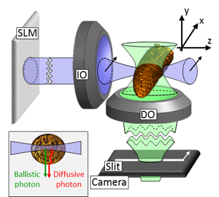
Separation of ballistic and diffusive fluorescence photons |
Sketch of the experimental setup
|
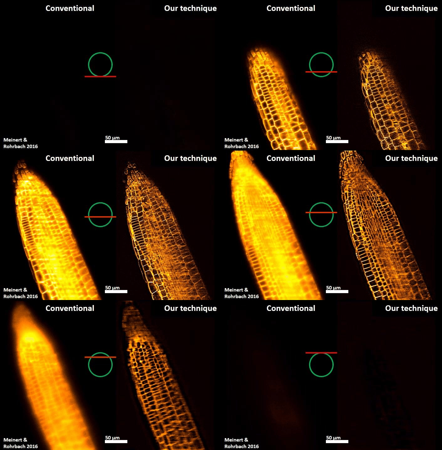
Arabidopsis Root Tip imaged by Light-Sheet_Microscopy. (Left) Illumination with Gaussian Beam,
conventional detection, and no post-processing (Right) Illumination with Bessel Beam, line
confocal detection and separation of diffusive and ballistic photons by post-processing.

- Meinert T, Rohrbach A
Light-sheet microscopy with adaptive Bessel beams
2019 Biomed Opt Express, Band: 10, Nummer: 2, Seiten: 670 - 681 - Bhat A, Meinert T, Michiels R, Rohrbach A
Miniature scanning light-sheet illumination implemented in a conventional microscope
2018 Biomed Opt Express, Band: 9, Nummer: 9, Seiten: 4263 - 4274 - Gohn-Kreuz C, Rohrbach A
Light needles in scattering media using self-reconstructing beams and the STED principle
2017 Optica, Band: 4, Nummer: 9, Seiten: 1134 - 1142 - Meinert T, Gutwein B-A, Rohrbach A
Light-sheet microscopy in a glass capillary – Feedback holographic control for illumination beam correction
2016 Opt Lett, Band: 42, Nummer: 2, Seiten: 350 - 353 - Meinert T, Tietz O, Palme K, Rohrbach A
Separation of ballistic and diffusive fluorescence photons in confocal Light-Sheet Microscopy of Arabidopsis roots
2016 Sci Rep-uk, Band: 6, Seite: 30378 - Gohn-Kreuz C, Rohrbach A
Light-sheet generation in inhomogeneous media using self-reconstructing beams and the STED-principle
2016 Opt Express, Band: 24, Nummer: 6, Seiten: 5855 - 5865 - Mousavi H, Gohn-Kreuz C, Rohrbach A
Feedback phase correction of Bessel beams in confocal line light-sheet microscopy – a simulation study
2013 Appl Optics, Band: 52, Nummer: 23, Seiten: 5835 - 5842 - Fahrbach F, Gurchenkov V, Alessandri K, Nassoy P, Rohrbach A
Light-sheet microscopy in thick media using scanned Bessel beams and two-photon fluorescence excitation
2013 Opt Express, Band: 21, Nummer: 11, Seiten: 13824 - 13839 - Fahrbach F, Gurchenkov V, Alessandri K, Nassoy P, Rohrbach A
Self-reconstructing sectioned Bessel beams offer submicron optical sectioning for large fields of view in light-sheet microscopy
2013 Opt Express, Band: 21, Nummer: 9, Seiten: 11425 - 11440 - Fahrbach F, Rohrbach A
Propagation stability of self-reconstructing Bessel beams enables contrast-enhanced imaging in thick media 2012 Nat Commun, Band: 3, Seite: 632 - F. O. Fahrbach, A. Rohrbach
A line scanned light-sheet microscope with phase shaped self-reconstructing beams
2010 Optics Express, Band: 18, Nummer: 23, Seiten: 24229 - 24244 - F. O. Fahrbach, P. Simon, A. Rohrbach
Microscopy with self-reconstructing beams
2010 Nature Photonics, Band: 4, Seiten: 780 - 785 - Rohrbach A
Artifacts resulting from imaging in scattering media: a theoretical prediction.
2009 Opt Lett, Band: 34, Nummer: 19, Seiten: 3041 - 3043


